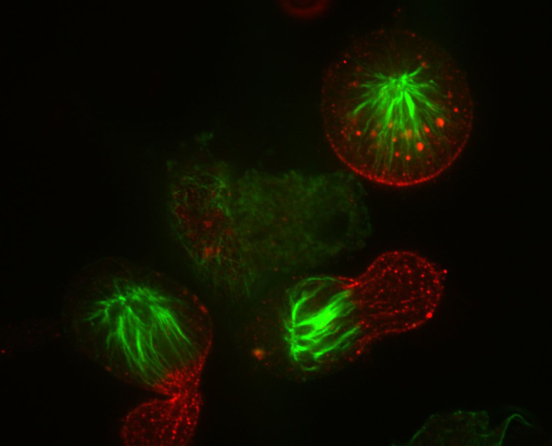
HeLa cells were accumulated in monopolar mitosis using a 12 hr treatment of the kinesin-5 inhibitor S-trityl-l-cysteine and forced into cytokinesis by adding the potent Cdk1 inhibitor purvalanol A for 15 min. The microtubules (green) were not growing and shrinking. Aurora B (red), which functions in attaching microtubules to the centromere, localizes at the gap region between the cortex and the microtubule plus ends. Cells were fixed with MeOH on ice for 3 min, and stained with a primary antibody against Auruoa B and FITC-DM1a antibody against microtubules. Secondary antibody was Alexa 594 for Aurora B. A single section was collected with a spinning disk microscope on a Nikon TE-2000 with a 1.3 NA 100X objective. Images were collected with an Orca ER CCD camera. Lasers and filters: Innova 70C Spectrum 3 watt Laser, 488 Laser/ATOF 525/50, and 568 Laser/ATOF 605/52.
Biological Process: Cytokinesis
Author: Hu, Chi-Kuo
Source: The Cell: An Image Library