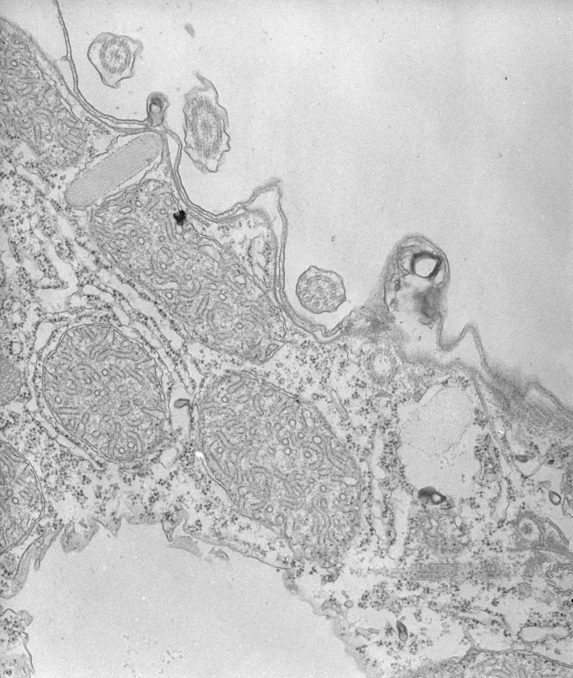
High resolution image of mucocysts which have a herringbone pattern of subunits and are surrounded by a membrane that fuses with the plasma membrane. When the mucocyst content is extruded the content expands (see Hausmann, Protistologica 8:401-412, 1972 for details. TEM taken on 8/12/67 by R. Allen with Philips 200 operating at 60kV. Neg. 19,200X. The raw film was scanned with an Epson Perfection V750 Pro. This image is best used for quantitative analysis.
Biological Process: Cortical granule exocytosis, Cortical organization
Standard glutaraldehyde fixation followed by osmium tetroxide, dehydrated in alcohol and embedded in an epoxy resin. Microtome sections prepared at approximately 75nm thickness. Additional information available at (http://www5.pbrc.hawaii.edu/allen/).
Author: Richard Allen (University of Hawaii)
Source: The Cell: An Image Library