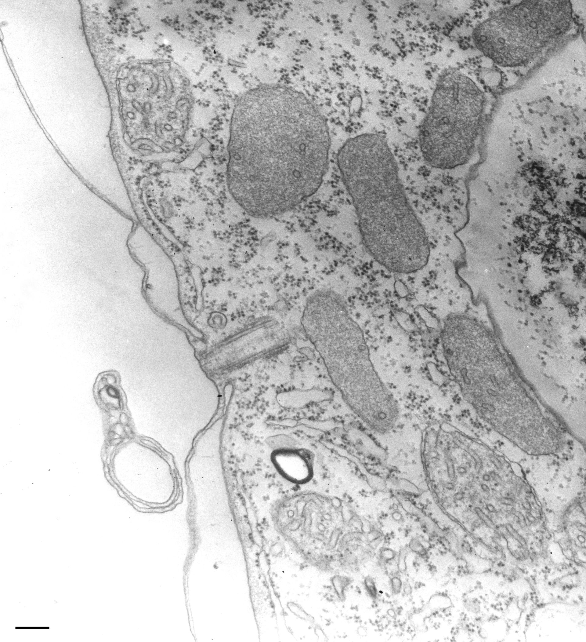
A particularly large accumulation of peroxisomes each surrounded by a single membrane and containing a few short tubules in an otherwise granular matrix. TEM taken on 8/9/65 by R. Allen with RCA EMU3F operating at 50kV. Neg. 12,700X. Bar = 0.2?m. The negative was printed to paper and the image was scanned to Photoshop. This digitized image is available for qualitative analysis. Additional information available at (http://www5.pbrc.hawaii.edu/allen/).
Biological Process: Cytoplasm organization
Standard glutaraldehyde fixation followed by osmium tetroxide, dehydrated in alcohol and embedded in an epoxy resin. Microtome sections prepared at approximately 75nm thickness. Additional information available at (http://www5.pbrc.hawaii.edu/allen/).
Author: Richard Allen (University of Hawaii)
Source: The Cell: An Image Library